


What is the Retina?
The retina is the light-sensitive nerve tissue lining the back of the eye (“the film in the camera”).
The retina is nourished by blood flow, which provides nutrients and oxygen that nerve cells need. A healthy, intact retina is the key to clear vision.
What is a retinal vein occlusion (BRVO / CRVO)?
A retinal vein occlusion means that a vein in the retinal circulation has become blocked. Retinal veins carry blood from the retina back to the heart for re-circulation.
If one of these veins becomes blocked a pressure build-up causes the walls of the vein to leak blood and excess fluid into the retina. When this fluid collects in the macula (the central area of the retina responsible for central vision), vision becomes blurry.
There are two types of retinal vein occlusions:
- Branch Retinal Vein Occlusion (BRVO): it happens when a smaller branch of the main retinal vein is blocked, damaging the area of the retina drained by this branch. Visual loss can happen but usually not as severely as a blockage of the main retinal vein (see below).
- Central Retinal Vein Occlusion (CRVO): it is caused by a blockage of the main retinal vein resulting in poor blood flow throughout the whole retina which can translate to severe vision loss.
What are the symptoms?
The most common symptom is vision loss or blurring in part or all of one eye. The symptoms may happen suddenly or become worse over several hours or days. Sometimes there is a sudden and complete loss of vision.
Pain in the eye can occur as a complication of a central retinal vein occlusion. This is caused by excessive eye pressure called neovascular glaucoma. This normally occurs a few months after a severe CRVO which has gone untreated.
Who is at risk?
Retinal vein occlusions are more common in:
- People over 65 years of age
- High blood pressure
- High cholesterol levels
- Diabetes
- People who smoke
- Glaucoma
- Some blood disorders
How is the condition diagnosed?
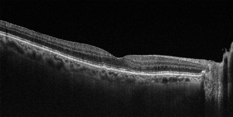
Dr Hilford will undertake a dilated examination of your eyes and tests such as Optical Coherence Tomography (OCT) and/or fluorescein angiography will be required.
What are the treatment options?
Dr Hilford will discuss the treatment options that are available which include intravitreal injections and/or laser treatment.